Cdc42 Pull-Down Activation Assay Kit
Cat. # 80701
Introduction
A. Background
Small GTPases are a super-family of cellular signaling regulators. Cdc42 belongs to the Rho sub-family of GTPases that regulate cell motility, cell division, and gene transcription. GTP binding increases the activity of Cdc42, and the hydrolysis of GTP to GDP renders it inactive.
Currently the activation of Cdc42 proteins is assayed with the binding of GTP-bound Cdc42 to the p21-binding domain (PBD) of p21-activated protein kinase (PAK). This method is based on the observation that the active, GTP-bound Cdc42 (and Rac) could bind to the PBD of PAK. However, the reproducibility of this method is poor. This is partially due to the relatively quick hydrolysis of GTP to GDP during the assay procedure, and the low binding affinity of PBD to Cdc42-GTP.
The Cdc42 Activation Assay Kit is based on the configuration-specific monoclonal antibody that specifically recognizes Cdc42-GTP, but not Cdc42-GDP. Given the high affinity of monoclonal antibodies to their antigens, the activation assay could be performed in a much shorter time. This assay provides the reliable results with consistent reproducibility.
B. Assay Principle
The Cdc42 Activation Assay Kit uses configuration-specific anti-Cdc42-GTP Mouse monoclonal antibody to measure Cdc42-GTP levels in cell extracts or in vitro GTPγS loading Cdc42 activation assays. Anti-Cdc42-GTP mouse monoclonal antibody is first incubated with cell lysates containing Cdc42-GTP. Next, the GTP-bound Cdc42 is pulled down by protein A/G agarose. Finally, the precipitated Cdc42-GTP is detected through immunoblot analysis using Anti-Cdc42 Rabbit Polyclonal Antibody.
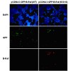
The anti-Cdc42-GTP monoclonal antibody can also be used to monitor the activation of Cdc42 in cells and in tissues by immunohistochemistry.
C. Kit Compo
nents
1. Anti-Cdc42-GTP Mouse Monoclonal Antibody (Cat. # 26905): One vial – 35 µL (1 mg/ml) in PBS, pH 7.4, containing 50% glycerol. This antibody specifically recognizes Cdc42-GTP from all vertebrates.
2. Protein A/G Agarose (Cat. # 30301): One vial – 600 µL of 50% slurry.
3. 5X Assay/Lysis Buffer (Cat. # 30302): One bottle – 30 mL of 250 mM Tris-HCl, pH 8, 750 mM NaCl, 50 mM MgCl2, 5 mM EDTA, 5% Triton X-100.
4. Anti-Cdc42 Rabbit Polyclonal Antibody (Cat. # 21010): One vial – 50 µL (1 mg/mL) in PBS, pH 7.4, contained 50% glycerol.
5. 100X GTPγS (Cat. # 30303): One vial – 50 µl at 10 mM, use 5 µL of GTPγS for GTP-labeling of 0.5 mL of cell lysate.
6. 100X GDP (Cat. # 30304): One vial – 50 µl at 100 mM, use 5 µL of GDP for GDP-labeling of 0.5 mL of cell lysate.
7. HRP-Goat Anti-Rabbit IgG (Cat. #29002): 50 µL (0.4 mg/mL) in PBS, pH 7.4, contained 50% glycerol.
D. Materials Needed but Not Supplied
1. Stimulated and non-stimulated cell lysates
2. Protease inhibitors
3. 4 °C tube rocker or shaker
4. 0.5 M EDTA at pH 8.0
5. 1.0 M MgCl2
6. 2X reducing SDS-PAGE sample buffer
7. Electrophoresis and immunoblotting systems
8. Immunoblotting wash buffer such as TBST (10 mM Tris-HCl, pH 7.4, 0.15 M NaCl, 0.05% Tween-20)
9. Immunoblotting blocking buffer (TBST containing 5% Non-fat Dry Milk or 3% BSA)
10. ECL Detection Reagents
E. Example Results
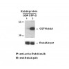
The following figure demonstrates example results seen with the Cdc42 Activation Assay Kit. For reference only.

Cdc42 Activation Assay. MEF cells were treated with (lane 2) or without (lane 1) EGF. Cell lysates were incubated with an anti- Cdc42-GTP mo
noclonal antibody (Cat. # 26905) (top panel). The precipitated active Cdc42 was immunoblotted with an anti-Cdc42 rabbit polyclo
nal antibody (Cat. # 21010). The bottom panel shows the Western blot with anti-Cdc42 of the cell lysates used (5% of that used in the top panel).
Assay Procedure
A. Reagent Preparation
1X Assay/Lysis Buffer: Mix the 5X Stock (Cat. # 30301) briefly and dilute to 1X in deio
nized water. Just prior to usage, add protease inhibitors such as 1 mM PMSF, 10 µg/mL leupeptin, or 10 µg/mL aprotinin.
B. Sample Preparation
Adherent Cells
1. Culture cells (one 10-cm plate, ~107 cells) to approximately 80-90% confluence. Stimulate the cells with activator or inhibitor as desired.
2. Aspirate the culture media and wash twice with ice-cold PBS.
3. Completely remove the final PBS wash and add ice-cold 1X Assay/Lysis Buffer (See Reagent Preparation) to the cells (0.5-1 mL per 10 cm tissue culture plate).
4. Place the culture plates on ice for 10-20 minutes.
5. Detach the cells from the plates by scraping with a cell scraper.
6. Transfer the lysates to appropriate size tubes and place on ice.
7. If nuclear lysis occurs, the cell lysates may become viscous and difficult to pipette. If this occurs, lysates can be passed through a 27½-gauge syringe needle 3-4 times to shear the genomic DNA.
8. Clear the lysates by centrifuging at 12,000 x g and 4°C for 10 minutes.
9. Collect the supernatant and store the sample (~1-2 mg of total protein) on ice for immediate use, or snap freeze and store at -70°C for future use.
Adherent Cells
1. Culture cells and stimulate with activator or inhibitor as desired.
2. Perform a cell count and then pellet the cells through centrifugation.
3. Aspirate the culture media and wash twice with ice-cold PBS.
4. Completely remove the final PBS wash and add ice-cold 1X Assay/Lysis Buffer (See Reagent Preparation) to the cell pellet (0.5-1 mL per 107 cells).
5. Lyse the cells by repeated pipetting.
6. Transfer the lysates to appropriate size tubes and place them on ice.
7. If nuclear lysis occurs, the cell lysates may become viscous and difficult to pipette. If this occurs, lysates can be passed through a 27½-gauge syringe needle 3-4 times to shear the genomic DNA.
8. Clear the lysates by centrifuging at 12,000 x g and 4°C for 10 minutes.
9. Collect the supernatant and store sample on ice for immediate use, or snap freeze and store at -70°C for future use.
C. In vitro GTPγS/GDP Protein for Positive and Negative co
ntrols
Note: In vivo stimulation of cells will activate approximately 10% of the available Cdc42, whereas in vitro GTPγS protein loading will activate nearly 90% of Cdc42.
1. Aliquot 0.5 mL of cell extract (or 1 µg of purified Cdc42 protein) into two microcentrifuge tubes.
2. To each tube, add 20 µL of 0.5 M EDTA (final concentration of 20 mM).
3. Positive control: add 5 µL of 100 X GTPγS (Cat. # 30302) to the 1st tube
4. Negative control: add 5 µL of 100 X GDP (Cat. # 30304) to the 2nd tube.
5. Incubate both tubes at 30°C for 30 minutes with agitation.
6. Stop loading by placing the tubes on ice and adding 32.5 µL of 1 M MgCl2 (final concentration of 60 mM).
D. Affinity Precipitation of Activated G Protein
1. Aliquot 0.5-1 mL of cell lysates (about 1 mg of total cellular protein) to a microcentrifuge tube.
2. Adjust the volume to 1 mL with 1X Assay/Lysis Buffer (See Reagent Preparation).
3. Add 1 µL anti-Cdc42-GTP antibody (Cat. # 26905).
4. Prepare the protein A/G Agarose bead slurry (Cat. # 30301) by resuspending through vertexing or titrating.
5. Quickly add 20 µL of resuspended bead slurry to above tube.
6. Incubate the tube at 4°C for 1 hour with gentle agitation.
7. Pellet the beads through centrifugation at 5,000 x g for 1 min.
8. Aspirate and discard the supernatant (making sure not to disturb or remove the bead pellet.
9. Wash the beads 3 times with 0.5 mL of 1X Assay/Lysis Buffer, centrifuging and aspirating each time.
10. After the third wash, pellet the beads through centrifugation and carefully remove all the supernatant.
11. Resuspend the bead pellet in 20 µL of 2X reducing SDS- PAGE sample buffer.
12. Boil the sample for 5 minutes.
13. Centrifuge it at 5,000 x g for 10 seconds.
E. Western Blot Analysis
1. Load 15 µL/well of pull-down supernatant to a polyacrylamide gel (17%). It is recommended to include a pre-stained MW standard (as an indicator of a successful transfer in step 3 below).
2. Perform SDS-PAGE following the manufacturer’s instructions.
3. Transfer the gel proteins to a PVDF or nitrocellulose membrane following the manufacturer’s instructions.
Note: Steps 4-11 are at room temperature with agitation
4. Following electroblotting, immerse the PVDF membrane in 100% Methanol for 15 seconds, and then allow it to dry at room temperature for 5 minutes.
Note: If Nitrocellulose is used instead of PVDF, step 4 Should be skipped.
5. Block the membrane with 5% non-fat dry milk or 3% BSA in TBST for 1 hr at room temperature with constant agitation.
6. Wash the blotted membrane three times with TBST, 5 minutes each time.
7. Incubate the membrane with Anti-Cdc42 Rabbit Polyclonal Antibody (Cat. # 21010), which has been freshly diluted 1: 50~500 (depending on the amount of Cdc42 proteins in your sample) in 5% non-fat dry milk or 3% BSA in TBST, for 1-2 hr at room temperature with constant agitation or at 4°C overnight.
8. Wash the blotted membrane three times with TBST, 5 minutes each time.
9. Incubate the membrane with a secondary antibody (Cat. # 29002), which is freshly diluted 1: 1000 in 5% non-fat dry milk or 3% BSA in TBST, for 1 hr at room temperature with constant agitation.
10. Wash the blotted membrane three times with TBST, 5 minutes each time.
11. Use the detection method of your choice such as ECL.
Cdc42 活性检测试剂盒/现货费斯德
Cdc42 活性检测试剂盒/现货费斯德
Cdc42 活性检测试剂盒/现货费斯德
本页产品地址:http://www.geilan.com/sell/show-9377690.html


 免责声明:以上所展示的[80701 Cdc42 活性检测试剂盒/现货费斯德]信息由会员[武汉费斯德生物科技有限公司]自行提供,内容的真实性、准确性和合法性由发布会员负责。